
LAB #12 - Marchantia polymorpha
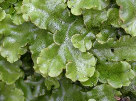
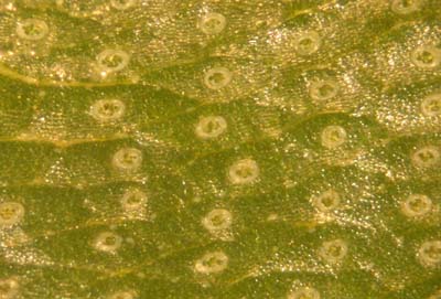 Surface View:
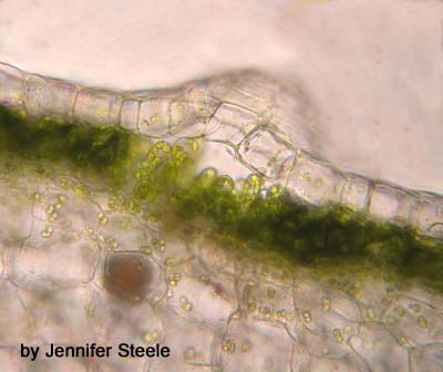
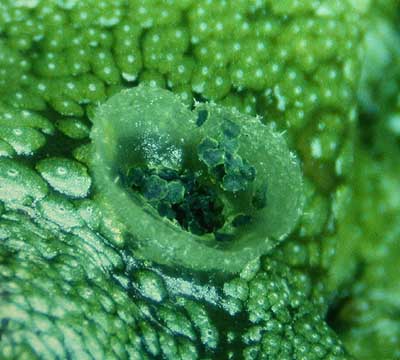
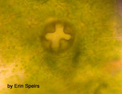 A closer look at a pore.
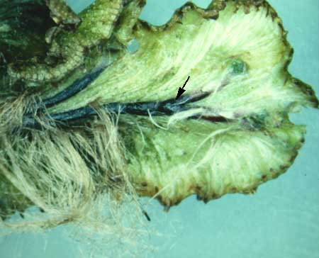
This shows you what the bottom surface of the thallus looks like. Note a scale indicated with an arrow.
Thallus cross-section:
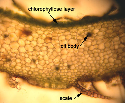
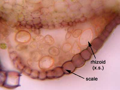
Cross-section through the lower side of the thallus showing a section through a scale and rhizoids in cross-section.
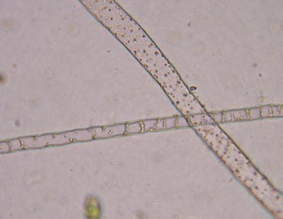 Rhizoids:
REPRODUCTIVE STRUCTURES:
Gemmae (Asexual Reproduction):
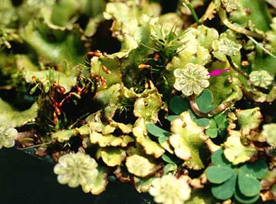
You can see the gemma cups (indicated with an arrow). These cups produce numerous small structures called gemmae which can grow into whole new plants.

 Close-up of a gemma cup.
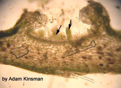
This is a longitudinal section through a gemma cup (arrows point to gemmae).
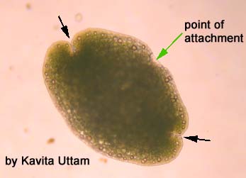
The two black arrows on the picture of gemma below indicate the growing points, from which new thalli will grow.
Antheridiophore:
Note the antheridiophore indicated with the pink arrow.
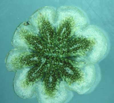
Thisis a look at the top of the antheridial disk.
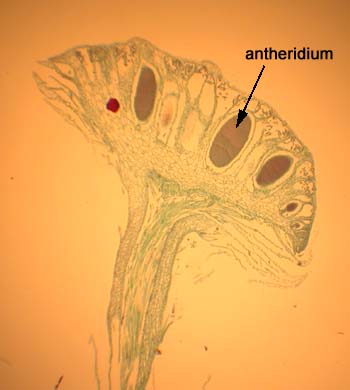
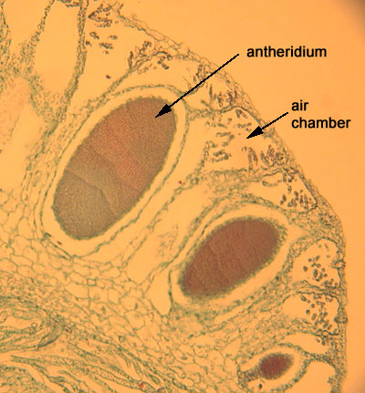
This is a picture of the longitudinal section through an antheridiophore.
The antheridia are immersed in the disk (top part).
Here is close-up of antheridia in a preparedslide.
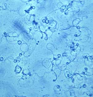 Sperm!
Archegoniophore:
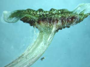
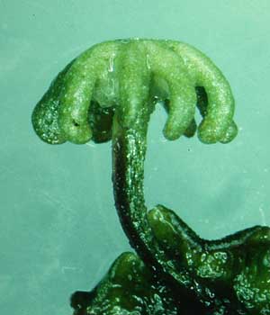
This is a young archegoniophore.
The longitudinal section through the top of the archegoniophore shows the archegonia (A), each with an egg or zygote within.
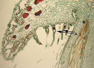
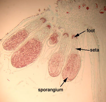
The zygote develops into a sporophyte. Here is a young sporophyte:
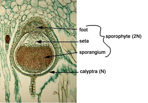
This longitudinal lection shows the sporophytes hanging down.
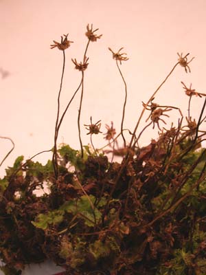
Here is what the archegoniophore look like after the spores have been released.