The microbial world
The living world extends far beyond that which our senses have evolved to interpret. Indeed, many have argued that the world of plants and animals that make up our everyday experience is just a thin veneer over a much larger and more variable microbial world. This microbial world is teeming with diversity that completely escaped our notice until a Dutch draper took his homemade microscope and looked into a drop of water in the late 1600s. Anton van Leeuwenhoek discovered a hidden biosphere that was neither plant nor animal, a discovery that modern scientific methods based on DNA sequencing has shown to have completely changed the face of biodiversity. Life is not primarily divided between plants and animals, but rather Bacteria, Archaea, and Eukaryotes. Plants, animals, and fungi are just three of the many lineages of Eukaryotes: all Bacteria and Archaea, and most Eukaryotes are microbial, and for most of the history of the planet all life was microbial. We are recent, and probably temporary guests on a microbial planet.
Portraits of Microbes
Art has drawn inspiration from nature from the first cave paintings to the present. Microbial life are part of the diversity of nature, but can they inspire art? Aren’t microbes just little round balls? They may be good at biochemistry, but are not animals and plants special because they have evolved unique and amazing diversities of physical form that capture our imagination? The answer, it seems, depends on how good is your microscope. When viewed up close, microbes are far from a boring collection of squiggling dots - microscopy can transport us into a parallel world of stunning complexity where microbes play all the leading roles. This world is abstract and far from our everyday experience, but it can be very engaging once you get inside.
Indeed, the abstract nature of the microbial world makes it difficult for microbiologists to engage non-experts beyond discussing the diseases some microbes cause. We hope that by illustrating intriguing microbial forms using metal, wood, and large-scale photographs, the pieces form portraits and landscapes from the alien world in which we live in a way that is more approachable. The work is not necessarily intended to be scientifically informative, but rather to contrast images from the microbial world with their presentation in the context of traditional art (e.g. huge gold picture frames). The images may clash with our expectations, but the work is simply art inspired by nature, just nature we can’t see with the naked eye.
What are these microbes and where do they come from?
Nearly all the microbes featured in this exhibit come from the most unlikely of places: the guts of wood-eating cockroaches and termites. Like other animals, these insects cannot digest cellulose (wood) and to survive they must harbour a diverse collection of Bacteria, Archaea, and single celled Eukaryotes (protists) in their gut, and these microbes carry out their digestion for them. Most of these species are found nowhere else on the planet. This arrangement, called ‘symbiosis’, or living together, gives the microbes a stable home and the host digestible food. Within the gut, various microbes also form symbioses with one another, so many bacteria live inside or on the surface of a protist, which can be seen in many of the pictures. Below, is short description of each piece, telling you what it is and a little bit about it, often beginning with a quote from its original description.
Spirotrichonympha operculum
“The species has an oval to pear-shaped cell body…Spirotrichonympha rotates slowly during its forward movement. The anterior flagella of one side of the body may extend towards the other side when sweeping forward.” Radek, 1997. Eur. J. Protistol.
Spirotrichonympha is at first glance a chaotic tangle of long filaments called flagella that project from the cell giving it the appearance of messy hair. But deep inside the cell is anything but chaotic. Like other complex protists, it is actually a highly organized structure with complex symmetry. In the case of Spirotrichonympha, the thousands of flagella are all organized along four helical bands that wind their way inside the cell from one end to the other. This image shows the tip of a Spirotrichonympha cell where the bands diverge and begin their spiral path through the cell.

Dinenympha
“The animal usually, remaining like its companions nearly stationary in position, writhes form side to side, shortens and widens or lengthens and contracts, and rotates in the long axis… a rapid waving motion, strikingly resembling the movement of flames” Leidy 1877. Proc. Acad. Nat. Sci. Philadelphia.
Dinenympha is an Oxymonad protist with eight flagella tightly bound to its body in a series of parallel helixes. It may not swim quickly, but its body pulsates with rapid convulsions due to its motile spine, or axostyle. The axostyle bends in a series of sharp kinks between straight segments that move down the length of the body in angular waves, lending it an unforgettable appearance.
Pseudotrichonympha cytoplasm (bubbles)
“Pseudotrichonympha is at once striking because of its great swimming power, exceeding that of any other protozoon of this termite. In living preparations it is a very pleasing sight to observe those animals gliding across the field of view, thrusting away with their anterior flagella the numerous wood particles and
other protozoa impeding their progress.” Cutler, 1919. Quart. J. Microsc. Sci.
Pseudotrichonympha is a large Parabasalian protist (not an animal), so large it can be quite delicate. Sometimes mishaps lead to interesting outcomes, and in this case the ‘skin’ or plasma membrane of a Pseudotrichonympha cell has been torn off cleanly as the cell was prepared for the scanning electron microscope. This gives us a rare 3D view inside the cell, where you can see an elaborate network of membrane vesicles (bubbles) and cytoskeletal connection (wormy squiggles), as well as the tubes through which the flagella would emerge in an intact cell.
Synderella glycocalyx (the hole)
“In Snyderella the nuclei have migrated into the deeper portions of the body where they are distributed without regularity and seem to have no connection with the blepharoplasts during the period of vegetative activity… Spirochaetes are almost always present in large numbers, each attached by one end to that part of the surface which is free of flagella.” Kirby, 1929. Univ. Calif. publ. zool.
The glycocalyx is a layer on the outer surface of many protists. It is a dense network of carbohydrates (long chains of sugars) that the cell exudes to protect its membrane from drying or damage. It can also make the cell sticky so they group together or form associations with other cells, like these bacterial symbionts. This image shows a small but perfect hole in the glycocalyx of the Parabasalian Snyderella.
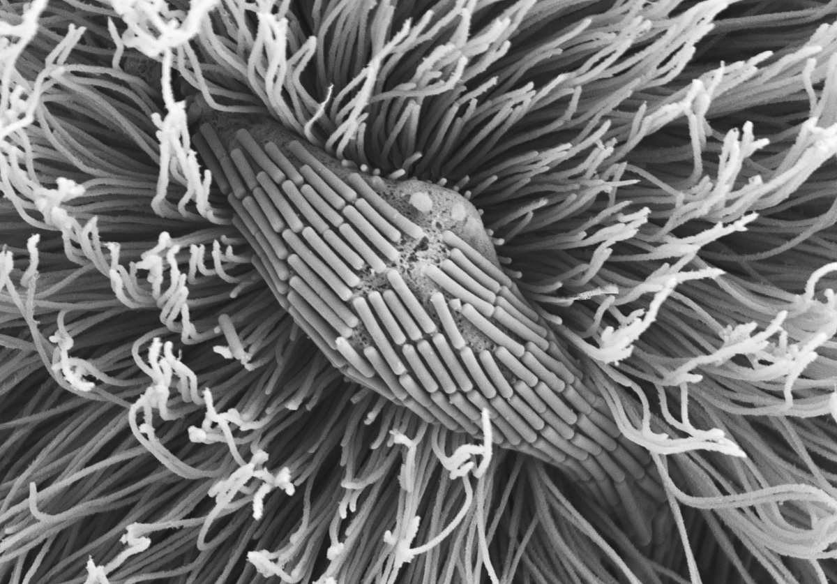
Trichonympha flagellar bands
“The most remarkable character of Trichonympha is its wonderful cloak of vibrating cils. No other animal of which I have any information has the appendages of such great length. They appear to emanate altogether from the summit of the head, and spread outward and backward enveloping the animals. And extending a considerable distance beyond its posterior extremity.” Leidy, 1881. The Parasites of the Termites.
Trichonympha is a large and very distinctive protist (not animal - in 1881 the distinction was vague) found in many termites and cockroaches. As Leidy notes above, the first thing one noticed about it is the flagella - a dense ‘cloak’ of long threadlike projects that wave and undulate in synchrony, transmitting waves of motion into the cytoplasm of the cell itself. This image shows a band of flagella adhered to one another to form overlapping sheets, presumably to aid in their synchronous undulations.
Calonympha axostyle
“This by Miss Dr. Anna Foà previously described flagellate belongs to the most beautiful and perhaps most complicated shapes nature has created in the group of protozoa; again and again the eye of the observer becomes drawn to the structures that are contained here in a single, if multi-nucleate, cell and endeavors to unravel them, certainly the success is not a complete one.” Janicki, 1915. Zeitschrift fur wissenschaftliche zoologie.
Calonympha is a Parabasalian flagellate with a large number of nuclei, the compartment where DNA is maintained. We usually think of cells having a single nucleus and genome, but in the evolution of Calonympha the entire complex including the nucleus, genome, flagella, and a cytoskeletal ‘spine’ called the axostyle has been massively replicated, giving rise to a cell with hundreds of each. In this image a bundle of axostyles emerges from the bottom of the cell.
Devescovina
“In the living flagellate the small rods often appear as double-contoured/double-outlined, strongly light refracting formations and one gets an impression of their exceptional delicateness and perishability: at longer observation of the living creatures the striatur/striped structure deforms and becomes disconnected from the (body) surface in the form of irregularly arranged finest rods.” Janicki, 1915. Zeitschrift fur wissenschaftliche zoologie.
Devescovina is an Parabasalian flagellate that is completely covered in a single layer of long, thin bacterial cells all oriented along the length of the slender Devescovina cell. These cells are visible in light microscopy, but their nature was not obvious. We now know they are living symbionts, attached to the surface of the protist presumably for some mutual advantage. Devescovina always has exactly four flagella, three that protrude forward, and one special one that extends to the rear. This rear-facing flagellum is broad and thick, taking on the appearance of a band or ribbon that can curl up on itself. At the other end, the cell tapers into a fine point, still coated with bacteria right to the tip.
Devescovina
See above.
Cthulhu
“represented a monster of vaguely anthropoid outline, but with an octopus-like head whose face was a mass of feelers, a scaly, rubbery-looking body, prodigious claws on hind and fore feet, and long, narrow wings behind."
"Ph'nglui mglw'nafh C'thulhu R'lyeh wgah'nagl fhtagn," Lovecraft, 1928. The Call of Cthulhu.
Cthulhu is one of the most recently described new Parabasalia, having been found in 2013. It is a relatively small cell, but distinguished by an asymmetrical tuft of 20 flagella that wave up and down synchronously. This gives it the appearance of a cephalopod, leading to its name after Cthulhu (said to be impossible to pronounce, but something like Ke-thoo-loo), the cephalopod-headed demon from HP Lovecraft. Taxonomy is often thought to be a bit stogy, but names like these are more memorable and also fun - in this case Cthulhu attracted the attention of Lovecraft fans, some media, and even has a song written about it. While this Cthulhu may not strike fear into your heart, its discovery helps us understand the natural diversity of termite symbionts.
Trichonympha split
“unless one has really seen this overwhelming mass of squirming, wriggling, undulating protozoa which greet the eye when a termite's intestinal content is viewed under the microscope, one cannot form any conception whatever of the immense difficulty envolved in attempting to study in detail any of them except the very large and dominant genus Trichonympha.” Cleveland, 1925. Biol. Bull.
As with the skinned Pseudotrichonympha, this large and delicate Trichonympha cell has split in half during processing, giving us another view inside a cell. The outside is orderly and symmetrical, but just below the surface is a complex confusion of vesicles and a dense network of fibers and tubes that makes up the cytoskeleton.
Oxymonas
“none is more unique than the development of an anterior proboscis capable of extension and retraction… In its function the proboscis appears to replace, at least in part, that of the flagella… ultimately, to lead to the entire elimination of flagella.” Kofoid & Swezy, 1926. Univ. Calif. publ. zool.
Oxymonas is, surprisingly, a member of the oxymonad lineage. Unlike most termite gut flagellates, Oxymonas likes to attach to the gut wall by way of a long proboscis, or extension of the cytoskeleton and cytoplasm made up of a long, rigid helix of cytoskeletal tubules and ending with a sticky cup that attaches to the animal. The whole proboscis can be retracted if the cell is disturbed. This Oxymonas, like many others, is covered in a patchwork of thin bacteria of various lengths, some of which are exceedingly tiny even by bacterial standards.
Saccinobaculus orbiting Barbulonympha
“It is difficult to give a word picture of the movement…. It moves almost constantly, and its most usual movement may be illustrated, somewhat crudely perhaps, by comparing it with a snake running in a bag…” Cleveland, 1934. Mem. Am. Acad. Arts Sci.
The name Saccinobaculus is indeed taken from the Huron for “Snake-in-a-bag” and this is an apt description because it has a long flexible ‘spine’ or axostyle that twists and bends inside the cell with astonishing rapidity, giving it the appearance of a wriggling bag. The axostyle is actually a wide band of microscopic tubules that rolls into a ball, or flattens out, or contracts as the tubules slide against one another at the molecular level. Saccinobaculus is only found in the cockroach Cryptocercus. This small Saccinobaculus is resting on a very large Barbulonympha cell whose surface makes up the entire background.
Barbulonympha apex
“The food of Barbulanympha, in common with the other protozoa that live in Cryptocercus, consists of wood particles which its host has eaten... The shape of the body resembles that of an acorn removed from its cup. Flagella arise only from the short, pointed, anterior portion. Forward movement, owing to the large body with a small flagellated area, is slower than that of most…” Cleveland, 1934. Mem. Am. Acad. Arts Sci.
Anyone that has eaten a rum ball, a chocolate treat popular at christmas, can identify with Barbulonympha. It is a giant (by microbial standards - the background of the central picture of Saccinobaculus is a small portion of a giant Barbulonympha cell) Parabasalian protist that is completely covered with a layer of identical rod-shaped bacterial cells. Its spherical shape and pattern of tiny “sprinkles” looks delicious, but Barbulonympha is not filled with chocolate and spirits, but rather with wood, which it eats and digests. At one end of this ball, Barbulonympha also has two symmetrical tufts of flagella separated by a thin bridge of its bacterial-covered surface, shown here.
Eucomonympha
“contains so many particles of wood that it frequently gives the appearance, when viewed at a low magnification, of a small bundle or collection of wood particles moving on its own accord in a somewhat haphazard manner.” Cleveland, 1934. Mem. Am. Acad. Arts Sci.
Eucomonympha is a large Parabasalian protist that is totally covered with up to 40,000 flagella. Its body is one cell, but is made of two separate compartments, giving it the appearance of a short, fat snow man. In addition to its flagella, its surface is also home to long thin bacterial symbionts called ‘spriochetes’. As Cleveland notes, Eucomonympha eats very large pieces of wood (hundreds of times larger than many of the other protists in the gut), and gorges itself until it seems it must burst. But it seldom does, even though it seems a delicate bag of sharp splinters.
Staurojoenina
“When first seen moving actively among the mass of flagellates and particles of wood removed from the intestine of the termite, Staurojoenina attracts attention at once by its agility and singular structure. The animal is in constant motion and the flagella continue beating until life is gone. The four bundles of flagella beat actively and independently. The animal is thus jerked from side to side but withal rowed forward, thrusting aside its lesser associates.” Kirby, 1926. Univ. Calif. publ. zool.
Staurojoenina resembles Barbulonympha in many ways (the two are actually pretty closely related), but rather than two tufts of flagella it has four. It is also covered in bacteria, which divide on the surface to form amazing patterns that resemble water currents or lines on a topographical map. These bacteria make tight physical connections to the protist, and at once place in the cell the lines of dividing bacteria converge (see on the lower left of the cell), forcing them to pile up and ultimately fall off.
Bacterial epibionts
"the lowest stage of the protist kingdom", as different from higher life as “a hydra was from a vertebrate...a simple alga from a palm” Haeckel, 1866. Generelle Morphologie der Organismen.
These bacteria actually live on the surface of a protist, Calonympha. The surface of Calonympha looks bare at a glance, but looking up close you see it is really an ecosystem unto itself, covered with a diversity of bacteria. The bacteria are attached to the surface of the cell, which is a sticky network of carbohydrates called a ‘glycocalyx’, which in some cases seems to almost swallow the bacterial cells.
Trichonympha ectoplasm
“The intestinal contents resemble a thick milky fluid, the great consistency of which is due to the vast numbers of protozoans which it contains, along with minute debris of wood particles. Through this mass the trichonymphs move with considerable rapidity, using the mobile anterior portion of the body to clear a pathway”. Kofoid & Swezy, 1919. Univ. Calif. publ. zool.
The anterior half of Trichonympha is where its flagella are organized. Though they seem to emerge at the surface of the cell, they actually emerge deep within the cell, and travel through tubes and folds to the surrounding water. This zone of the cell looks different and was called ‘ectoplasm’ by early microscopists. Here you can see rows of flagella emerging from folds of the ectoplasm.
Bacterial landscape
"the comparatively little-modified descendants of whatever single form of life appeared on earth" Copeland, 1956. The classification of lower organisms.
While bacterial diversity is not generally associated with shape and size, here a variety of both are visible in a landscape reminiscent of a coral reef. Long thin corkscrews are spirochetes, while others are rods or spheres of various sizes and identities. The community of bacteria in a termite gut is very unusual and highly adapted to the environment, which lacks oxygen (it would be toxic to bacteria and protists alike) and is rich in some nutrients and sparse in others. Many bacteria now appear to play a role in metabolising nitrogen in exchange for energy, because wood is nitrogen poor but energy rich.
Trichonympha quadriptych (1 of 4)
See below.
Trichonympha quadriptych (2 of 4)
See below.
Trichonympha quadriptych (3 of 4)
See below.
Trichonympha quadriptych (4 of 4)
"The arrangement of the long cilia, clothing the body, reminded [me] of the nymphs in a recent spectacular drama, in which they appeared with their nakedness barely concealed by long cords suspended from the shoulders, and this arrangement has suggested the name applied to the parasite." Leidy 1877. Proc. Acad. Nat. Sci. Philadelphia.
Trichonympha was the first of the many protists in termites to be observed by one of the fathers of protistology, Joseph Leidy. His name cast a mold for the naming of other large protists, many of which end in “-nympha”. What is less known, however, is that his name was inspired by a “spectacular drama” and more specifically some scantily clad ladies in this drama. Trichonympha is a Parabasalian protist, and one of the most widespread symbionts in termites and cockroachs, being found in hundreds of host species around the world. It is large and distinctive, covered in hundreds to thousands of flagella, the hair-like waving appendages usually used for swimming. These four pictures reconstruct a Trichonympha cell: on the left is the fanned ‘cap’ or operculum, the middle two show details of their thousands of elaborately organized flagella, and at the right is the posterior end of the cell, here covered with pine nut-shaped bacteria, where it actually eats particles of wood.